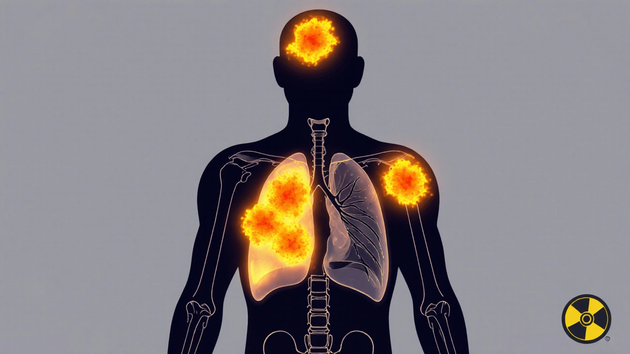
Why accurate cancer staging depends on the right imaging tool
Getting cancer staged correctly isn’t just a technical step-it changes everything. A misread scan can mean delayed treatment, unnecessary surgery, or missing a hidden spread. That’s why doctors don’t rely on one scan alone. They pick between PET-CT, MRI, and sometimes PET-MRI based on the cancer type, location, and what they’re trying to find. The goal? To know exactly how far the cancer has gone, where it’s hiding, and whether treatment is working. There’s no single best scan. Each has strengths, limits, and situations where it shines.
PET-CT: The workhorse of cancer staging
PET-CT became the standard in the early 2000s because it shows two things at once: where cancer is active and where it’s located in the body. It uses a sugar-based tracer, usually 18F-FDG, injected into the bloodstream. Cancer cells, which burn sugar faster than normal cells, light up on the scan. The CT part gives the detailed anatomy-like a map showing exactly which lymph node or organ is affected.
It’s fast: a full-body scan takes about 15 to 20 minutes. It’s widely available, even in smaller hospitals. For lung cancer, lymphoma, and colorectal cancer, PET-CT is often the first choice. A 2023 meta-analysis found it correctly identifies spread to lymph nodes in non-small cell lung cancer with 84% accuracy. But it’s not perfect. Some cancers, like prostate or certain liver tumors, don’t absorb the tracer well. And because it uses radiation-10 to 25 millisieverts per scan-it’s not ideal for young patients or those needing repeated scans.
MRI: Seeing soft tissue in high definition
MRI doesn’t use radiation. Instead, it uses powerful magnets and radio waves to create incredibly detailed images of soft tissues. This makes it the go-to for brain tumors, spinal cancers, liver lesions, and pelvic cancers like prostate or cervical cancer. A 3T MRI machine can show structures as small as half a millimeter. For prostate cancer, MRI detects tumors with 75% accuracy, compared to 62% for PSMA PET-CT in some studies.
Its real power comes in spotting subtle changes. After radiation therapy, it can tell the difference between scar tissue and returning cancer-a critical call. A 2023 review found MRI alone correctly identifies tumor recurrence in brain tumors about 70-80% of the time. But MRI is slow. A detailed scan can take 30 to 60 minutes. It’s also noisy, claustrophobic for some, and can’t be used if you have metal implants like pacemakers or certain surgical clips. Not every hospital has the right equipment or staff trained to read complex MRI scans.

PET-MRI: The high-end hybrid
PET-MRI combines the metabolic insight of PET with the soft-tissue detail of MRI in one machine. It was first approved in 2011 and has slowly grown in academic centers, especially in Europe and the U.S. It reduces radiation exposure by about half compared to PET-CT because it skips the CT scan. This makes it ideal for children, young adults, and patients needing long-term monitoring.
For certain cancers, it’s a game-changer. A 2023 study in RadioGraphics found PET-MRI changed treatment plans for nearly half of pancreatic cancer patients because it spotted small metastases PET-CT missed. In liver cancer, 68% of radiologists reported higher confidence in diagnosing lesions with PET-MRI than with PET-CT. For brain tumors, it outperforms both: accuracy in distinguishing recurrence from radiation damage jumps to 85-90% with PET-MRI, compared to 70-80% with MRI alone.
But it’s not for everyone. The machine costs over $3 million to install. Scans take 45 to 60 minutes. Motion during the scan-like breathing or fidgeting-can blur images. Only about 25% of U.S. cancer centers have one. And because it’s newer, interpretation requires extra training. Radiologists need an additional 3-6 months of specialized education beyond standard nuclear medicine or radiology training.
How doctors choose: It’s not one-size-fits-all
There’s no universal rule. The choice depends on the cancer and the question.
- For breast cancer: PET-CT is better at spotting early response to chemo before three cycles. MRI is better for checking the original tumor size and spread to the chest wall.
- For prostate cancer: PSMA PET-CT is great for finding distant spread, but MRI is still the gold standard for seeing if cancer is inside the prostate itself.
- For brain tumors: PET-MRI is becoming the preferred option because it tells you if a new growth is cancer returning or just inflammation from treatment.
- For pediatric cancers: PET-MRI is increasingly used to cut radiation exposure over a child’s lifetime.
- For lymphoma: PET-CT remains the standard for staging and checking if treatment worked.
Dr. Richard L. Wahl from Johns Hopkins puts it simply: “PET-MRI’s superior soft tissue contrast makes it valuable for pelvic and brain tumors, but PET-CT remains the workhorse.” That’s because availability, speed, and cost still matter. A small community hospital can’t justify a $4 million PET-MRI machine when PET-CT does the job for most patients.
Cost, access, and the future of imaging
PET-CT costs between $1,600 and $2,300 in the U.S. PET-MRI? $2,500 to $3,500. That 50% price difference isn’t just about the machine-it’s the staff, training, maintenance, and longer scan times. A 2022 survey found 45% of cancer centers struggled with reimbursement and workflow when adding PET-MRI.
Still, adoption is growing. The global PET-MRI market is projected to grow nearly 19% per year through 2030. New systems like Siemens’ BioMatrix 600, cleared in early 2024, cut scan times to just 6 minutes for a full-body scan. That’s a big step forward.
What’s next? Artificial intelligence is starting to help. Algorithms now analyze PET-MRI scans to predict how a tumor will respond to treatment before it even shrinks. The NCI’s PREDICT trial is testing whether AI can use imaging patterns to pick the best drug for each patient. And new tracers-like those targeting PSMA in prostate cancer or somatostatin receptors in neuroendocrine tumors-are making PET scans more specific than ever.
What patients should know
If you’re facing cancer imaging, ask: Why this scan? Not every cancer needs a PET-CT. Not every case needs an MRI. Sometimes, a simple ultrasound or X-ray is enough. If your doctor recommends a PET-MRI, ask if it’s because your cancer type is known to benefit from it-or if it’s because your center has the machine and wants to use it.
Also, know your radiation exposure. If you’ve had multiple CT scans in the past, ask if MRI or PET-MRI could reduce your total dose. If you’re young or planning to have children, radiation matters. If you have metal implants, MRI might not be an option.
There’s no perfect scan. But there’s a right one for your situation. The goal isn’t to get the fanciest machine-it’s to get the most accurate information with the least risk.
What’s next for oncologic imaging?
The future isn’t about replacing one tool with another. It’s about combining them smarter. Think of it like a team: MRI sees structure, PET sees activity, and AI connects the dots. By 2035, PET-MRI could be used in 25-30% of cases in major cancer centers, especially for complex or recurrent cancers.
For now, the most important thing is this: imaging isn’t magic. It’s a tool. And like any tool, its value depends on who’s using it-and why.


Written by Felix Greendale
View all posts by: Felix Greendale